Living in the era of break-through technologies, lots of people still suffer from undiagnosed and untreated eye diseases. AIRA retina analysis tool leverages AI capabilities to provide patients with reliable and cost-effective ophthalmological care.
Unfortunately, many people still experience lack of up-to-date medical treatment and ophthalmological patients are no exception. In 2022 there are at least 2.2 billion people with some kind of vision impairment. The good news is that in almost half of these cases there are still high chances of recuperation. Luckily, we now have a variety of powerful technologies able to make cutting-edge healthcare solutions.
AIRA (AI Retina Analyzing and Disease Diagnosis Tool) is one of such solutions. Our most skillful AI and machine learning specialists partnered with the leading ophthalmologists to deliver top-notch retina healthcare applications able to detect abnormalities in the eye structure.
In this article we will show what modern technologies can help enhance retina analysis and treatment and present to you the details of our AIRA project.
What are the consequences of inadequate eye diagnosis?
Healthy eyes are of crucial importance to our well-being so once an eye disease is left unnoticed or the treatment is not accurate, it can lead to visual distortion. In the worst scenario, a person can become completely blind.
In its turn, blindness will cause major difficulties with continuing day-to-day activities and will require expenditures on compensatory equipment and/or services of a medical attendant.
Quite an unpleasant perspective, to say the least. That’s why incorporating novel technologies is so important to achieve the most precise diagnosis results.
What are the challenges hindering proper eye diagnosis?
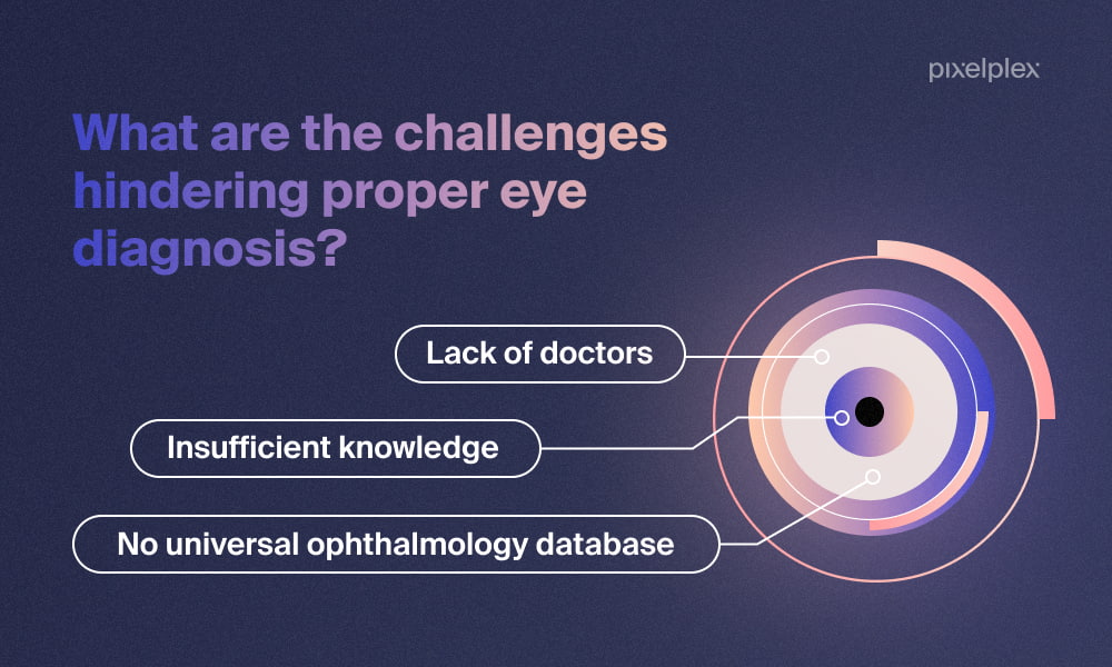
There are several difficulties in the traditional eye healthcare analysis approach that may badly affect the diagnosis outcome and lead to inadequate treatment. These include:
Insufficient knowledge
Academic time dedicated to ophthalmology training is relatively short, meaning that medical students have lots of complicated information to grasp on a tight schedule. Meanwhile, ophthalmology is one of the specializations that require both excellent surgical skills and deep clinical experience.
Lack of doctors
Ophthalmology is considered one of the most difficult and competitive medical specializations so it is no wonder that there are lots of vacant positions. It is estimated that the US will demand additional 72,500 ophthalmology specialists by 2029. Speaking of the global scale, there should be 14 million practicing optometrists to fulfill the demand for proper eye treatment. There were only 331,743 specialists worldwide in 2021.
The situation will get more complicated as the number of patients over age 65 will increase 42% by 2030, meaning each specialist will take extra load. This will inevitably lead to longer appointment waiting times and increased possibility of diagnosis mistakes due to the overwhelming amount of medical data a specialist will need to attend to.
No universal ophthalmology database
There is no comprehensive ophthalmology database where specialists could turn to check their diagnosis or learn some new information. Thus ophthalmologists have to rely on segmental texts and images from various sources that have no common standards and which can be prone to bias since they are mostly labeled manually.
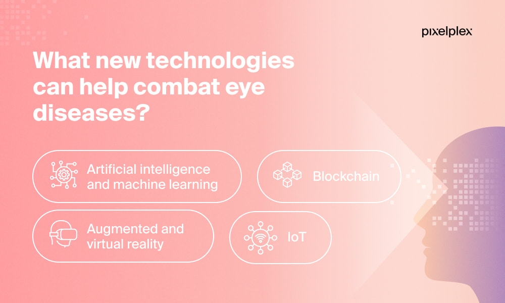
What new technologies can help combat eye diseases?
There are several cutting-edge technologies able to fight the aforementioned challenges and increase efficiency of eye disease diagnosis. They include artificial intelligence, machine learning, blockchain, and augmented and virtual reality.
Let’s take a look at what these technologies are exactly capable of.
Artificial intelligence and machine learning
When applied in ophthalmology, artificial intelligence (AI) and its subset, machine learning (ML) can process huge amounts of patients’ data in search of common patterns, such as symptoms. These technologies are extremely precise, so they can detect slightest details that a human specialist can overlook. It is especially beneficial for doctors who have started gaining experience just recently.
Assigning image processing to AI, rather than humans, significantly reduces time as the technology can “look through” a great amount of samples in a short time and doesn’t require any rest. This way, ophthalmologists can focus on more complex tasks and deliver diagnosis and treatment much faster than when they do manual scanning.
AI and ML-based solutions can also be trained to analyze patients’ medical history and lifestyle patterns such as environment, working conditions, and habits to predict the likelihood of developing an eye disease.
In addition, ophthalmologists can leverage Natural Language Processing (NLP) to process unstructured data from various sources: text, audio, and video, so that it may be later added to the universal database.

A Complete Guide to Healthcare Business Intelligence and Tips on How to Use It Effectively

When You Need Blockchain For Your Project and When It Is Not the Best Option
Meet CheckNFT.iO — an AI-powered solution able to analyze non-fungible tokens and detect fraudulent activities
Blockchain
Blockchain technology can serve as a great foundation for a universal ophthalmology base. Stored on blockchain, medical data can be easily accessible to lots of people, from practicing doctors to students.
This will enhance quality of diagnosis, treatment, education, and allow doctors effortlessly consult with colleagues from other locations in case they have any doubts or difficulties.
Since no change on the ledger can be done discreetly, there will be no possibility for fraudsters to tamper with the data. What’s more, eye specialists will be able to put timestamps on the medical records or pictures, making it easy to track progress and turn to interesting cases in a few clicks.
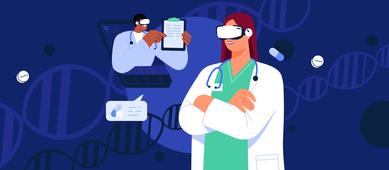
Augmented and virtual reality
Augmented (AR) and virtual (VR) reality technologies can be of much help in the education of next generation ophthalmologists. Students can use surgical and diagnostic training simulators based on VR and AR systems to get sufficient practice and hone their skills.
Besides, AR and VR can be used to spread public awareness about eye diseases. Specialists can imitate different eye abnormalities and suggest people experience how they feel with the help of a VR headset or an AR mask.
IoT
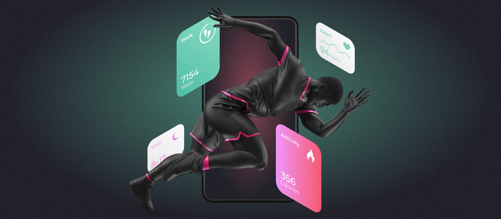
Internet of things (IoT) is one more technology able to help ophthalmologists. It is mostly used for remote patient monitoring and allows to automatically collect health metrics such as heart rate, blood pressure, and temperature.
This way doctors can track patients’ conditions not requiring in-person appointment. IoT remote monitoring is a perfect option for people who already experience visual distortions and have difficulties with commuting to a medical facility. Also, in case any metric seems suspicious, the IoT device can send alerts both to doctor and patient, and even suggest what measures to undertake.
Predictive Analytics in Healthcare: Use Cases and Key Benefits Explained
Artificial Intelligence Applications in Healthcare
Learn more about digital transformation in healthcare
AIRA: a solution by PixelPlex for diagnosing retinal diseases
Our AI development team has teamed up with experienced ophthalmologists to provide a high-tech solution for automated retinal abnormalities symptom identification and diagnosis.
Powered by AI, AIRA makes its conclusions based on a large dataset of retinal images, including both healthy samples and those showing signs of disease. The dataset comprises images with visual defects and of different quality, which allowed us to train AIRA on a much deeper level.
Data collection
The development team at AIRA was provided with a large dataset of retinal images with the image masks for the following symptoms, and morphological and anatomical features:
- Exudates;
- Hemorrhages;
- Microaneurysms;
- Disk;
- Cup;
- Drusen;
- Veins;
- Arteries.
To build a comprehensive dataset, the images were collected from different sources by using different cameras with varying qualities. The image quality varied depending upon the camera and the opacity of the eye structure. Some images also had minor visual artifacts adding to the diversity of data.
Data preprocessing
Many machine learning algorithms require data to be in a consistent and uniform format. Same is true for neural networks that require a fixed size for all inputs. All data images were hence converted to RGB format. They were normalized and resized if needed.
Data generation
When training deep learning networks, it is common practice to increase the size of available data by synthetically generating new data points. The dataset for AIRA was augmented by generating new images by using the following techniques:
- Affine transformations such as rotations, shift, skew etc.
- Change of color and quality by applying various filters like blurring, contrasting, hue, saturation, etc.
Training via U-Net
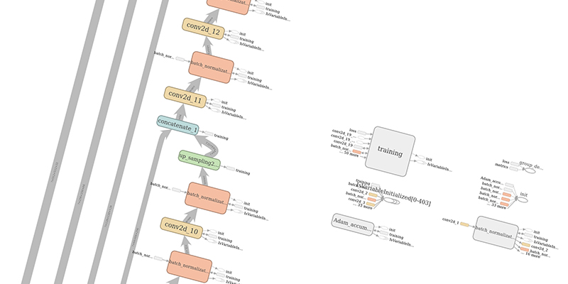
U-Net, derived from CNNs, has several contracting layers followed by expansion layers. Its architecture gives it a “U” like appearance and ensures feature maps learnt in the contracting layers are directly passed on to expansion layers used for reconstructing output segmented image maps.
PixelPex’s team developed a highly specialized deep learning model based on U-Net after several trials and extensive experimentation. The technical features in the final network architecture are the following:
- Activation function: ReLu;
- Using layers of batch normalization: + Method of increasing the dimension: Upsampling;
- Count of filters in the mid feature vector: 512;
- The dimension of the input/output layer: 512×512;
- Optimizer: Adam Accumulate;
- Missing data: Extrapolation by mirroring.
Implementation via the above parameters resulted in precise and accurate segmentation and prediction while using fewer training images. Integrating AIRA in a fundus camera would enable novice and inexperienced doctors to accurately and reliably diagnose retinal diseases. Skilled doctors can use AIRA’s platform to train a network using their own specialized images adding to the knowledge base.
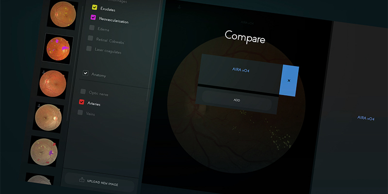
Example results
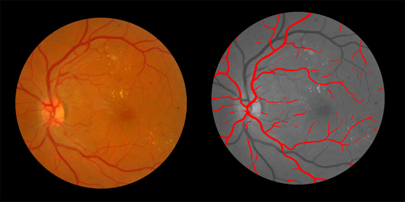
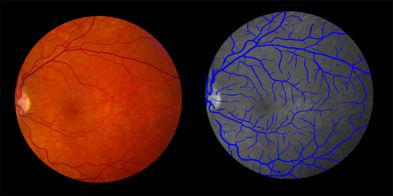
The advantage of employing U-Net is that each image is labeled pixel by pixel to identify various structures or anomalies present in the retina. Two broad categories identified by AIRA are anatomical structure or a symptom. Examples of the two different tasks are illustrated below. In each figure, the input image is shown on the left and the identified anatomy or symptom is shown in color on a gray-scaled background.
Identification of anatomical structure
Shown below are examples of arteries and veins identified by AIRA:
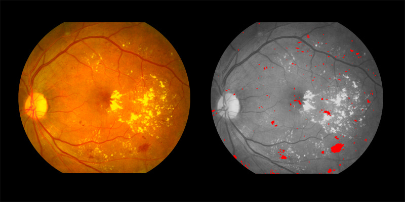
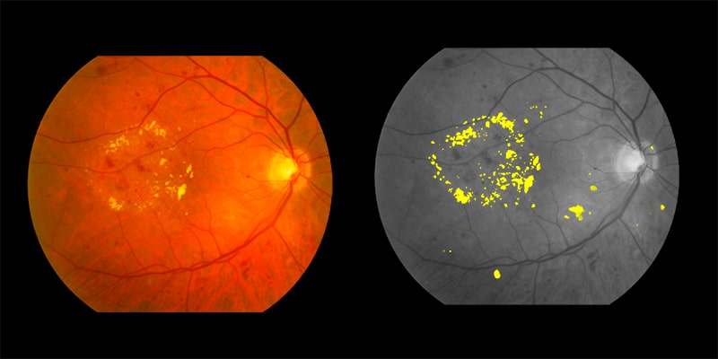
Symptoms identification
Two examples of symptoms pinpointed by AIRA are shown below:
Contribution to a universal knowledge base
AIRA has been trained on a set of retinal images making it a general platform for retinal disease diagnosis and aid doctors making an accurate diagnosis. Ophthalmologists can provide their own training images with masks for additional symptoms and anatomies to further specialize the system for other retinal pathologies. This would enhance the analysis and prediction of the neural network and broaden its domain to cater to a wider set of retinal pathologies.
Large numbers of people around the world do not have access to specialists despite having investigative tools like a fundus camera. AIRA is a valuable tool that can help medical staff make an initial assessment of retinal diseases in patients.
AIRA can also help monitor patients as the disease regresses and to define the difference in symptoms as percentages, which humans would find hard to do; and serve as an aid to making referrals to specialized doctors if needed.
Crafting a Top-Tier Health Monitoring App: Insights From PixelPlex's Real-World Project
The Role of AR and VR Technologies in Healthcare

The Synergy of Blockchain and Artificial Intelligence: Unlocking New Business Opportunities
Get acquainted with other game-changing healthcare projects we deployed
Conclusion
We are certain that advanced technologies such as AI, ML, blockchain, AR/VR, and IoT will gain more traction in the healthcare domain, boosting high-level diagnosis and treatment of patients.
Our team feels very inspired by the AIRA retina diagnosis solution since it has a massive potential to improve the way retinal diseases are analyzed and help more people get timely diagnosis and treatment.
Wish to enrich the healthcare tool pool with the next high-tech solution? We’d readily assist you with that. Drop us a line and let’s discuss your idea right away.